Supriono, Harijono Achmad
Division of Gastroenterology and Hepatology Department of Internal Medicine
Brawijaya University School of Medicine/ Dr. Saiful Anwar Hospital, Malang, East Java, Indonesia
Introduction: Imaging-guided percutaneous catheter drainage has evolved to become a first-line method to treat pancreatic pseudocysts. The indications for draining pseudocysts include presence of symptoms or complications and progressive enlargement. We report of 27 years old man with traumatic pseudocyst of pancreas who successful managed by ultrasound (US)-guided percutaneous catheter drainage.
Case report: The patient admitted with chief complaint abdominal enlargement since two months ago, gradually onset, and feel stabbing-like pain continuously. He also suffered from nausea and vomiting. He was alcoholism and history of traffic accident, his abdomen was hit by steer three months ago. On physical examination, the patient looked dyspneu, restlessness and abdominal enlargement (fig-1). The result of US was suspected large pancreatic pseudocyst with more than 27 cm in diameter (fig-2). We decided to perform percutaneous catheter drainage with US-guided (fig-3). The pseudocyst was containing more than 3 liter of hemorrhagic fluid. After underwent drainage, we performed CT-scan with the result was insertion the drain until the tail of the pancreatic duct (fig-4). The condition of patient became gradually better in the monitoring (fig-5).
Discussions: The large of pancreatic pseudocyst is a very rare case. The patient falls in critical condition, because the pseudocyst had progressive enlargement. The management of this patient was still challenges. US-guided percutaneous drainage was the first priority managed the patient to reduced compression of the abdomen.
Conclusion: Percutaneous catheter drainage in critical patient with large pancreatic pseudocyst should be performed, first minimally to reduced compression of the abdomen.

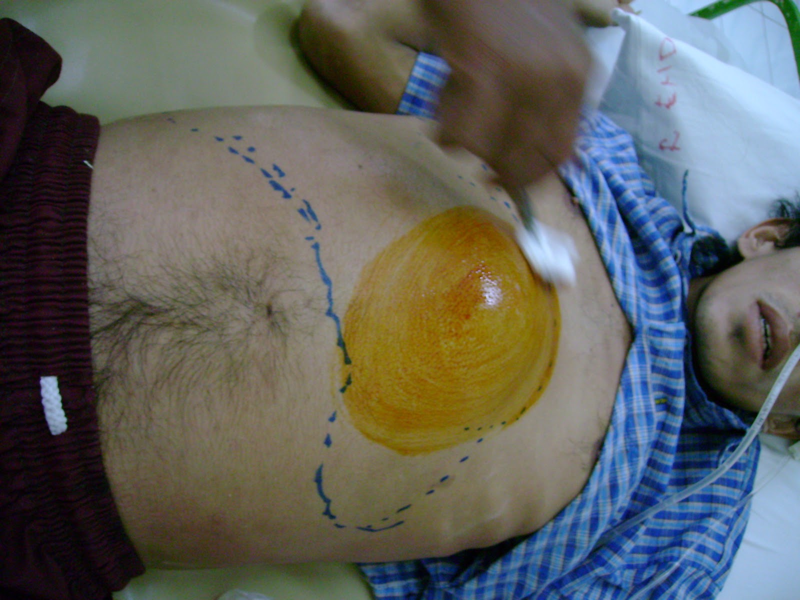


Fig-3


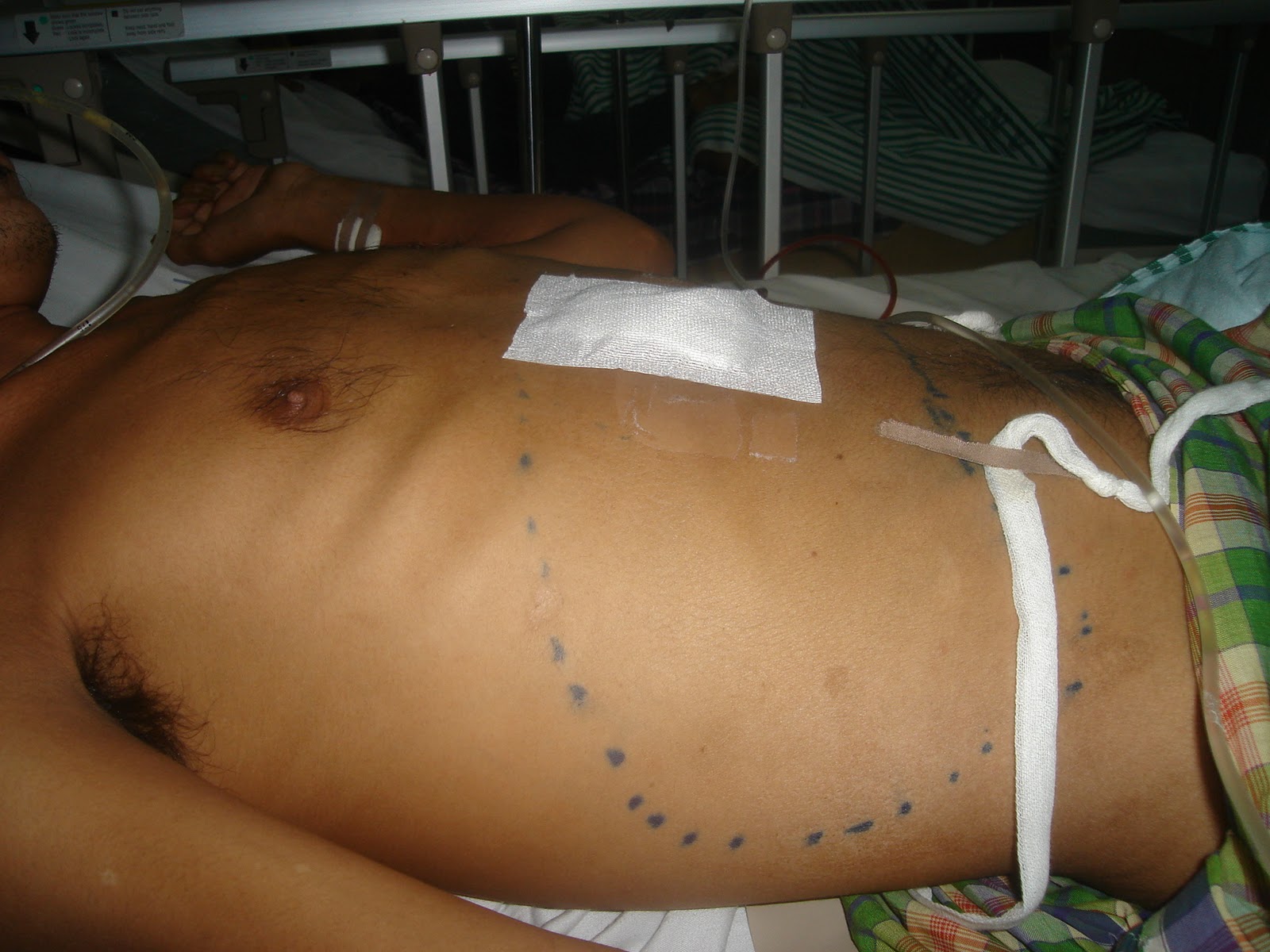
Fig-5
Tidak ada komentar:
Posting Komentar